In brief
Epidermal, dermal and mixed melasma describe pigment depth. Centrofacial, malar and mandibular describe where pigmentation appears on the face. These are different ways of describing melasma, not six competing diagnoses or a menu of treatments. DermNet: melasma.
If your notes say “mixed malar melasma,” the words answer two separate questions. They do not tell you, on their own, which procedure to choose or how much improvement to expect. This guide explains how to read those descriptions and what an assessment can—and cannot—establish.
For the separate question of whether a patch is melasma, a post-inflammatory mark or another form of pigmentation, start with melasma versus hyperpigmentation. Matching a photograph to a category is not a substitute for diagnosis.
First separate depth from location
Imagine a clinical note with two fields: suspected pigment depth and facial distribution. Completing one does not complete the other. “Malar” does not mean superficial, and “mixed” does not tell you which part of the face is involved.
The useful follow-up is not “Which type is the worst?” It is “What does this description tell us, how was it assessed, and would it change the plan?”
Epidermal, dermal and mixed: what the depth terms mean
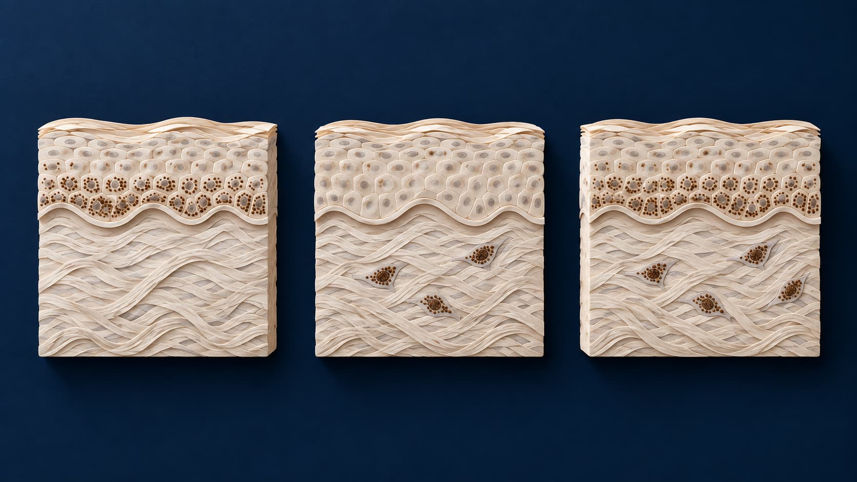
The epidermis is the outer skin layer; the dermis lies beneath it. The traditional classification concerns the level of increased pigment, rather than the darkness of a photograph.
| Term | What it describes | What the word alone does not establish |
|---|---|---|
| Epidermal | Increased pigment in the epidermis | A guaranteed quick response or a reason to select a peel |
| Dermal | A pigment component in the dermis | That treatment is pointless or must be more aggressive |
| Mixed | Epidermal and dermal pigment components | An exact proportion in each layer or a fixed treatment sequence |
These are established descriptive categories. They are not measurements in millimeters. DermNet: melasma classification.
Centrofacial, malar and mandibular: the facial map
The distribution terms describe recognizable patterns:
- Centrofacial: central facial areas, including the forehead, cheeks, nose and part of the upper-lip area.
- Malar: the cheeks and nose.
- Mandibular: the jawline and chin.
These names describe location, not a separate pigment layer. DermNet: facial patterns. A familiar distribution also does not prove that an unfamiliar patch is melasma; clinicians sometimes need to exclude another condition. AAD: melasma diagnosis.
What a Wood’s lamp and dermoscopy can add
A dermatologist often recognizes melasma through examination. A Wood’s lamp or dermatoscope may add information, rather than replace the clinical assessment. A biopsy is sometimes considered when another condition needs to be ruled out; it is not required for everyone with melasma. AAD: diagnosis and treatment.
Wood’s lamp: a change in contrast, not an automatic verdict
A Wood’s lamp uses a specialized light source. Pigment that becomes more conspicuous can support an epidermal component; lack of enhancement is traditionally associated with deeper pigment. Interpretation still matters. Products or residue on the skin can affect the appearance under the lamp, so tell the clinician what you have applied. Do not try to reproduce the examination with a household blacklight. DermNet: Wood lamp examination.
Dermoscopy: closer detail that still needs interpretation
A dermatoscope combines magnification and illumination to reveal skin structures more clearly. It is not simply a phone camera zoomed in on a dark patch. DermNet: dermoscopy.
Nor should either instrument be treated as an infallible pigment-depth scanner. In a 50-person cross-sectional study, Wood’s lamp and dermoscopic classifications showed only moderate agreement. The study did not compare either method with histology or reflectance confocal microscopy, so it cannot establish which tool measured the true depth more accurately. It illustrates a limitation of classification—not a reason to dismiss a useful examination. Navya and Pai, 2022.
Why color charts and online photos are not enough
Brown or gray tones can be clues for a clinician, but they are not a reliable home sorting test. You cannot infer an exact pigment layer from a screen or assume that a similar-looking patch has the same cause. Even clinical imaging is affected by its light source: an optical study found that the same reference colors looked different under different dermoscopes and lighting conditions. That study was about color reproduction, not a validated way to classify your melasma. Hanlon and colleagues, 2022.
A depth label is not a treatment recommendation
“Epidermal” does not automatically mean “have a peel,” and “dermal” does not mean “choose a stronger laser.” A plan also needs to account for skin tone, relevant triggers and the individual's circumstances. There is no single best treatment for everyone with melasma. AAD: individualized treatment.
Procedures can worsen pigmentation or cause unwanted lighter areas. Their possible benefits and risks need a separate explanation, not a shortcut from a category name. British Association of Dermatologists: melasma.
Similarly, the label does not guarantee clearance, establish a recovery time or predict that improvement will be permanent. Melasma may recur after treatment. “Mixed” is not a percentage score, and “dermal” is not a personal prognosis. Ask which findings actually support the expectations being discussed.
For broader context on matching a goal to a modality, see how facial treatments are selected. That overview is not a melasma-specific procedure recommendation.
Turn the terminology into useful questions
You do not need to arrive having chosen a type. Bring the wording from any earlier assessment and a short account of what you have tried and how your skin responded. Then ask:
- What was described? Was the term about depth, location or a different feature?
- What supports it? What did the examination show, and what remains uncertain?
- Would more testing change a decision? What specific question would a lamp, dermatoscope or biopsy help answer?
- What is a reasonable goal? What change would count as worthwhile, and how would we assess it?
- Why this approach? What alternatives are reasonable, including not having a procedure?
- What would make us reconsider? What risks, reactions or lack of progress would change the plan?
The point is to understand the reasoning, not to collect every available test. The AAD's consultation guidance emphasizes discussing realistic outcomes, alternatives, risks and recovery before agreeing to a cosmetic procedure.
Sources
Sources checked September 24, 2026 (Mexico City). Professional references explain general assessment, not the availability of a particular diagnostic device or treatment at Juvenalia Brío.
- DermNet. Melasma: classification and facial patterns.
- American Academy of Dermatology. Melasma: diagnosis and treatment.
- DermNet. Wood lamp skin examination.
- DermNet. Dermoscopy.
- Navya A, Pai V. Comparison of Dermoscope and Woods Lamp as a Tool to Study Melanin Depth in Melasma. Indian Dermatology Online Journal. 2022;13(3):366–369. doi:10.4103/idoj.idoj_245_21.
- Hanlon KL, Wei G, Correa-Selm L, Grichnik JM. Dermoscopy and skin imaging light sources: a comparison and review of spectral power distribution and color consistency. Journal of Biomedical Optics. 2022;27(8):080902. doi:10.1117/1.JBO.27.8.080902.
- British Association of Dermatologists. Melasma.
- American Academy of Dermatology. Questions before cosmetic treatment.